Contact Us
100 Cummings Center, Suite 407-P, Beverly, Massachusetts 01915, USA
+1 (978) 720-8044
Contact Us
100 Cummings Center, Suite 407-P, Beverly, Massachusetts 01915, USA
+1 (978) 720-8044

Explore Our Case Studies
Thrive Bioscience's suite of cell biology solutions empowers researchers by enhancing the quality and depth of data across a wide range of applications. Thrive systems enable researchers to push the boundaries of discovery with greater precision and efficiency. From tracking cell growth to documenting complex biological processes, our solutions deliver detailed, reproducible, data-driven insights crucial for advancing research.
Thrive Bioscience's instruments and software provide critical support for a variety of applications that require advanced live-cell imaging. Our products are designed to assist you in conducting:
Automated Cell Counting:
Accelerate and enhance the accuracy of cell counting, reducing variability and ensuring consistent results across experiments.
Growth Rate Analysis:
Track and analyze cell growth over time with high precision, providing valuable insights into cell behavior and culture conditions.
Data Logging and Documentation:
Automatically log and document all stages of the research process, creating a detailed, traceable record that supports reproducibility.
Confluence Measurement:
Accurately measure confluence to monitor cell proliferation and ensure optimal conditions for experimental success.
Accelerate and enhance the accuracy of cell counting, reducing variability and ensuring consistent results across experiments.
Track and analyze cell growth over time with high precision, providing valuable insights into cell behavior and culture conditions.
Case Studies
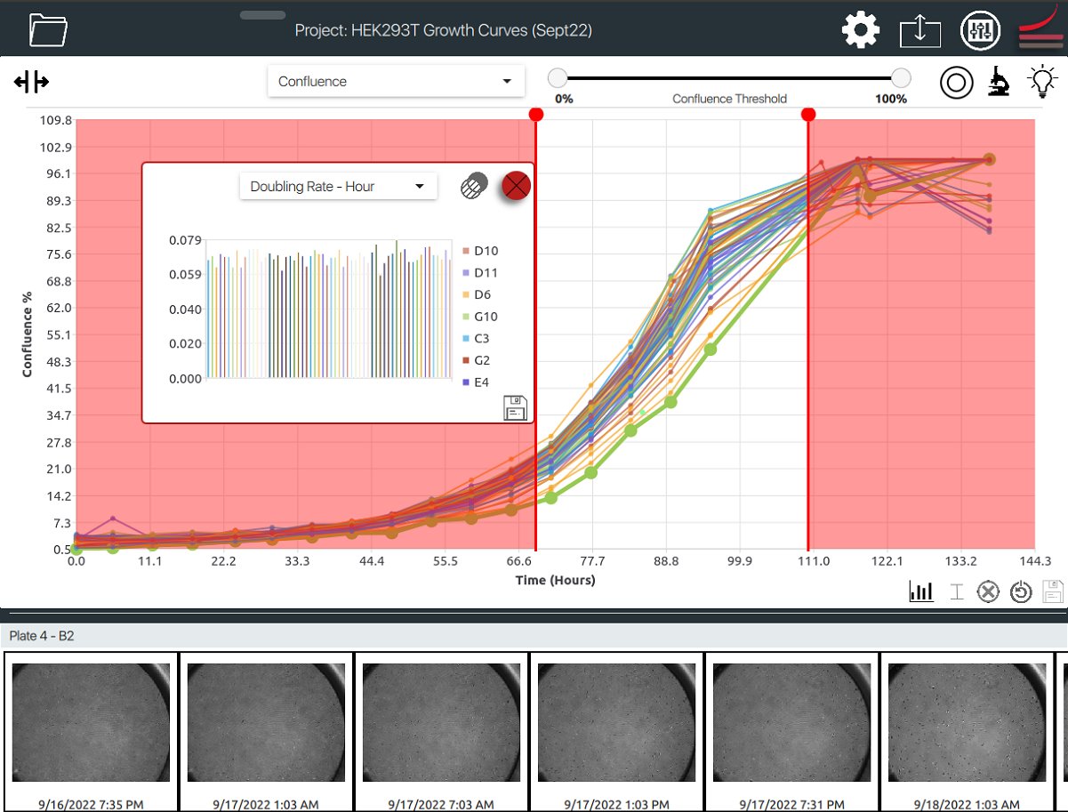
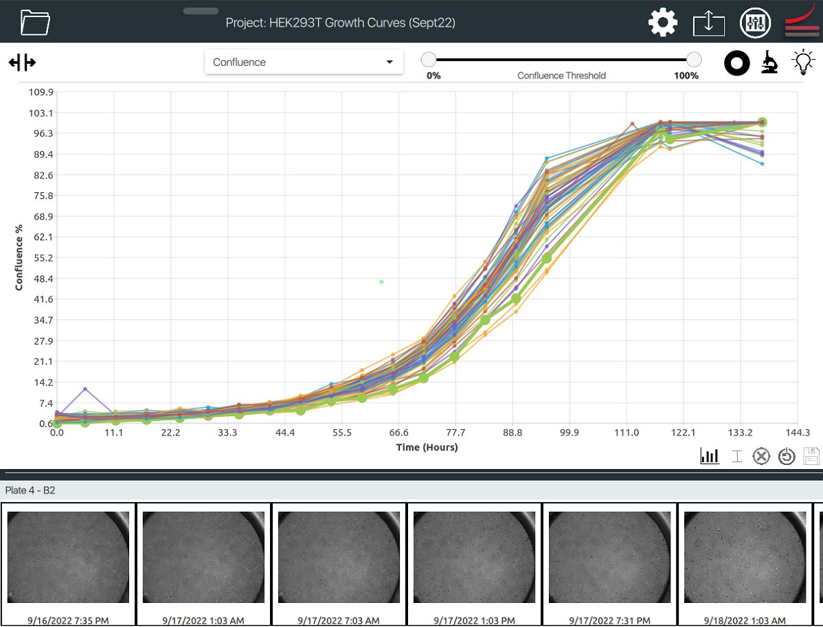
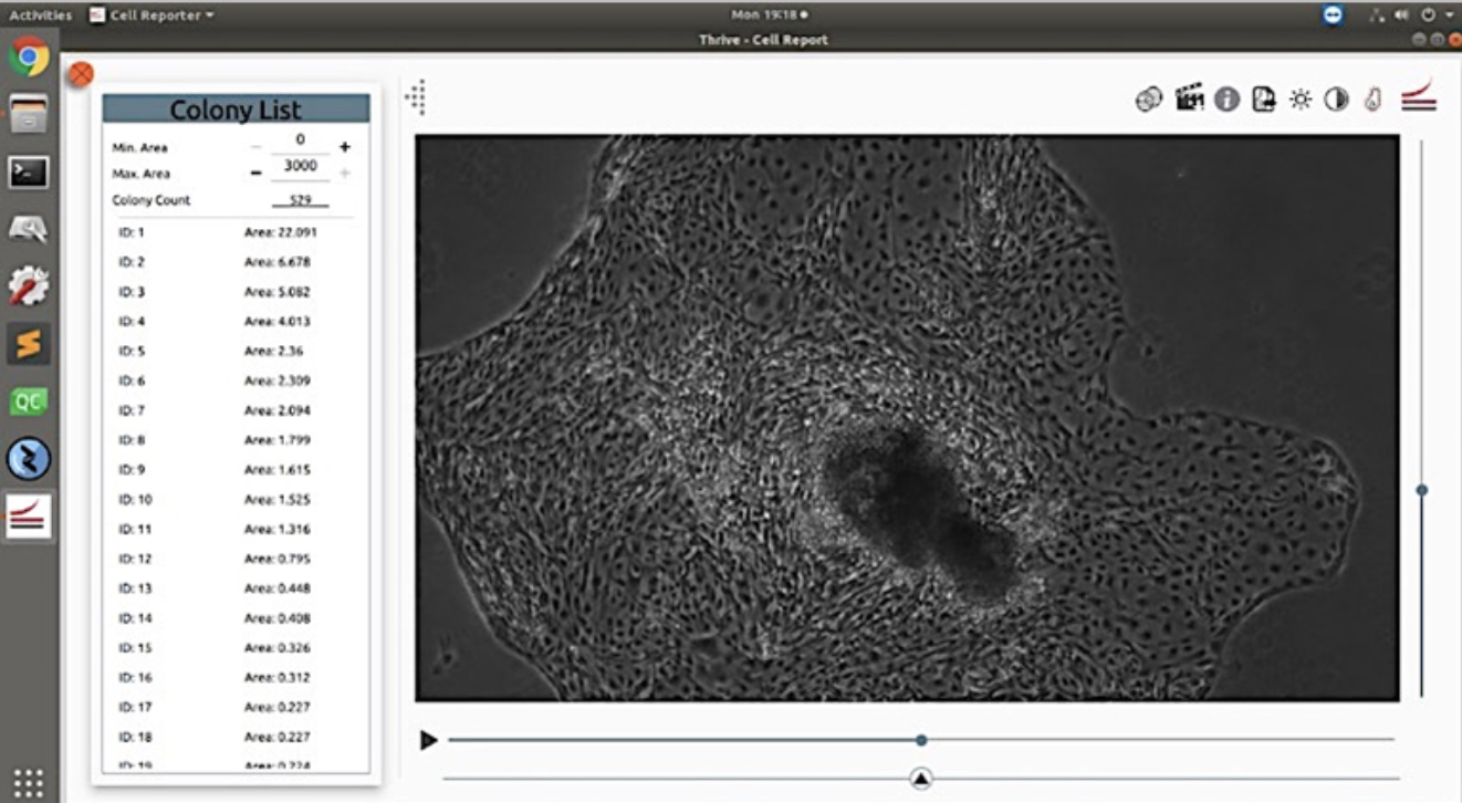
The CellAssist Systems improve the quality and documentation of cell cultures.
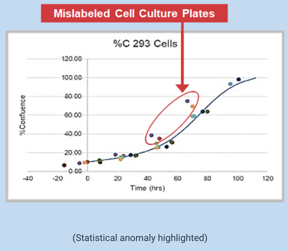
Problem: Current tools do not collect images and data along with tools that enable users to visualize outliers. Outliers are important because they may indicate problems in quality that require investigation.
Objective: Document and analyze images and data on cells over time to improve the quality and reproducibility of cell, tissue, and suspension cultures.
CellAssist Solution: The CellAssist Systems provide comparable data over time that allows for the identification of outliers. (In this example, upon further investigation, the customer found cases of mislabeling.)
CellAssist improves quality and increases yield by using data to grade and pick colonies.
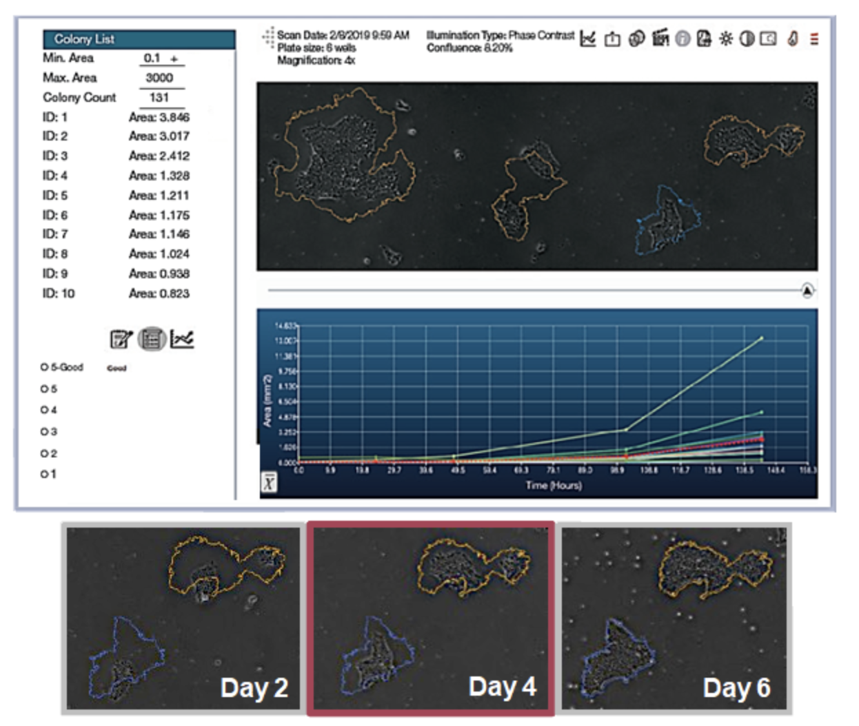
Problem: Gene editing transfection rates can be poor, so selecting monoclonal colonies for passaging is crucial.
Objective: Accurately determine whether a colony is monoclonal by capturing a series of time-lapse images with excellent registration across scans.
CellAssist Solution: The CellAssist Systems' time-lapse imaging enables the lineage of candidate colonies to be tracked to a single starting cell.
Morphological assays do not disturb cell growth
Problem: Differentiation of neuronal progenitor cells into glial cells was measured using fluorescence-based endpoint assays, which did not allow for further growth and study of the cells. An equivalent assay was needed based on imaging of live-cells.
Objective: Acquire high-resolution images that are comparable over time to study differentiation without dyes and/or probes that interfere with the cells and colonies.
CellAssist Solution: The CellAssist Systems' high-resolution stacks of images, with highly accurate registration across scans, enabled the characterization of cell morphology without disturbing the cells and colonies.
Optimization, Yield, and Cost Savings
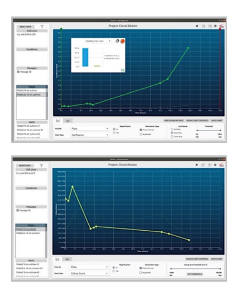
Problem: The coating of plates can profoundly affect the survivability and growth of colonies (in this case, gene-edited ones starting with very small quantities). Some coatings are also significantly more expensive.
Objective:
1. Coating Comparison: Evaluate and compare two different coatings by observing and characterizing the behavior of cells under each condition over a set period. This analysis will help determine which coating optimally supports cell health and activity.
2. Protocol Optimization: Develop, implement, and assess new protocols tailored to these coatings. By directly comparing the results, the most effective protocol and coating combination that enhances experimental outcomes can be identified.
CellAssist Solution: The CellAssist Systems' documentation, colony counting curves, and image examination characterize the number of colonies for each condition, and the results are collated and easily compared across conditions.
Elevate Your Research with Thrive Bioscience's Advanced Live-Cell Imaging Solutions
Thrive Bioscience's suite of advanced live-cell imaging applications is revolutionizing cell biology research by providing precision, efficiency, and deep insights across a range of applications. Our innovative solutions support the next generation of scientific breakthroughs, from stem cell research and drug discovery to wound healing and personalized medicine. By leveraging our cutting-edge technology, researchers can streamline workflows, enhance data quality, and drive meaningful results faster.
Ready to elevate your research capabilities?
Let's Talk.
Explore the CellAssist Systems designed to streamline researcher workflows, enhance data quality, and drive discovery with precision and efficiency.
View More
Connect with one of our expert staff to discuss how Thrive Bioscience technologies can enhance your cell biology research.
Contact Us
Learn more about our technologies and their use in advancing cell biology research.
RESOURCES
Application Note
The CellAssist 50 in BSL-3 Facilities
May 1st, 2025
View More
RESOURCES
Application Note
Assessment of Growth and Doubling Time Changes with Increasing Passages Utilizing CellAssist 50
April 1st, 2025
View More
RESOURCES
Application Note
Variability in Cell Confluency: Comparison of Human and CellAssist Assessments
March 1st, 2024
View More