Problem:
1.) The coating of plates can profoundly affect the survivability and growth of colonies (in this case gene edited ones starting with very small quantities).
2.) Some coatings are also significantly more expensive.
Objective:
1.) Coating Comparison: Evaluate and compare two different coatings by observing and characterizing the behavior of cells under each condition over a set period. This analysis will help determine which coating optimally supports cell health and activity.
2.) Protocol Optimization: Develop, implement, and assess new protocols tailored to these coatings. By directly comparing the results, identify the most effective protocol and coating combination that enhances experimental outcomes.
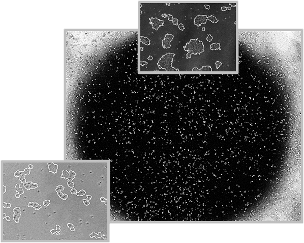
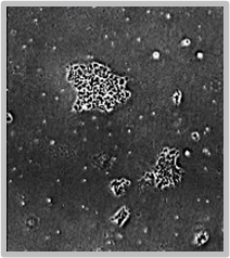
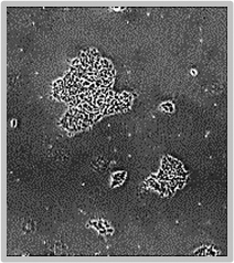
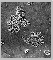
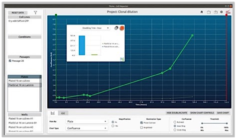
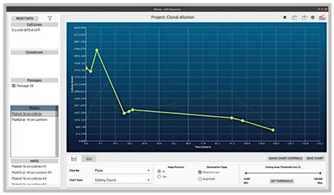
CellAssist Solution:
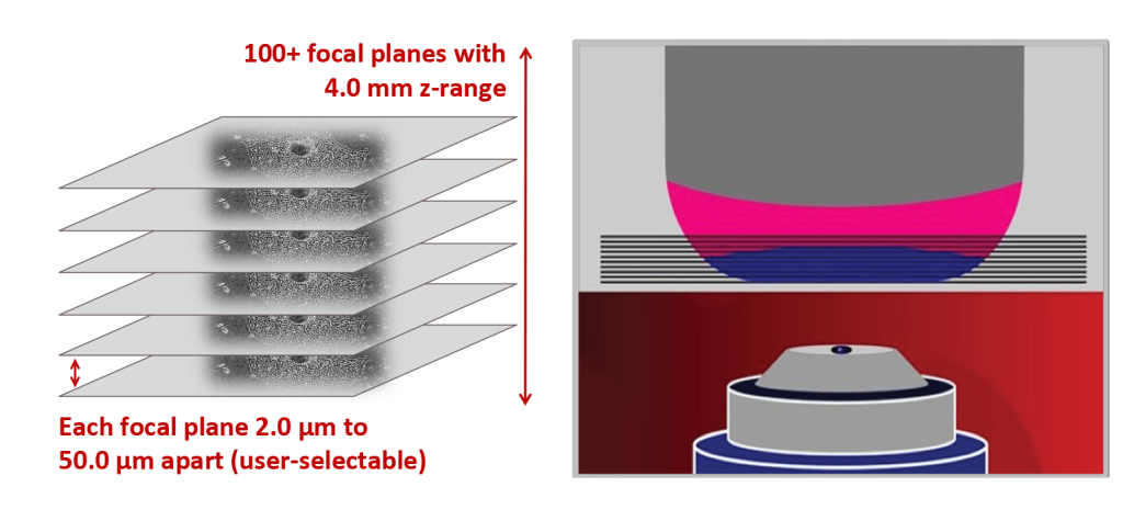
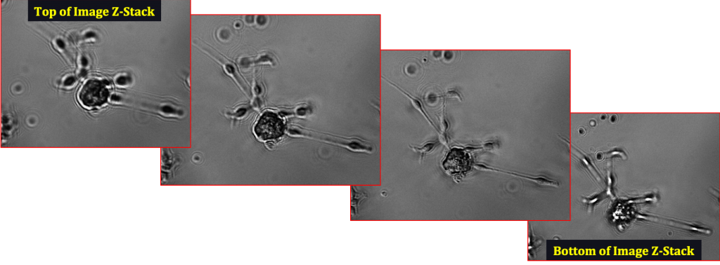
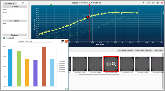
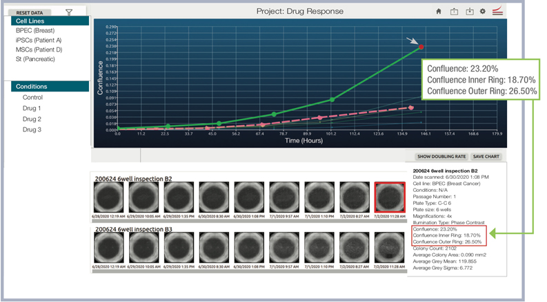
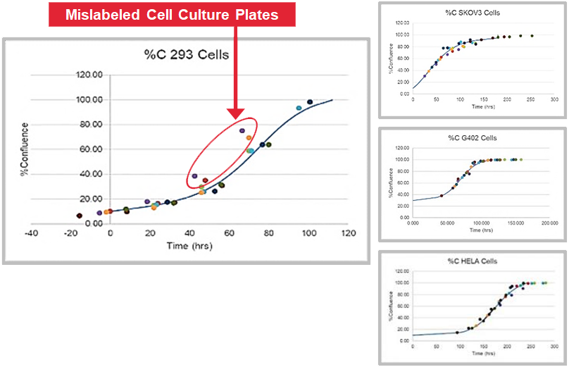
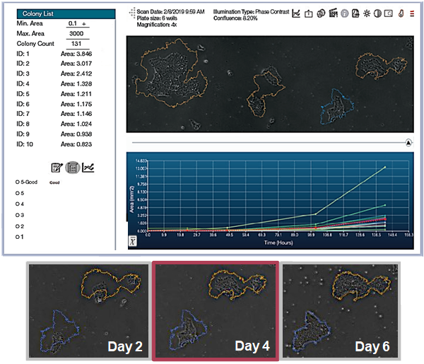
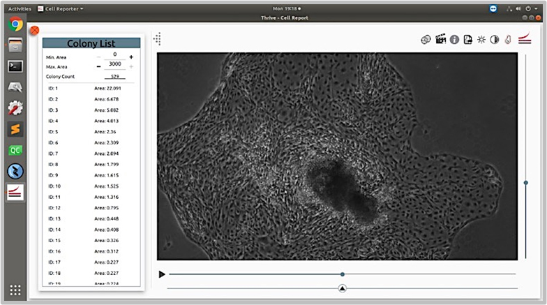
The CellAssist’s documentation, colony counting curves, and examination of images are used to characterize the number of colonies for each condition, and the results are collated and easily compared across conditions.