Contact Us
100 Cummings Center, Suite 407-P, Beverly, Massachusetts 01915, USA
+1 (978) 720-8044
google-site-verification=_llX3wt-_sILNZ4OKcVhHaUFsbcxl34KDwm75iPzM38
Contact Us
100 Cummings Center, Suite 407-P, Beverly, Massachusetts 01915, USA
+1 (978) 720-8044
Contact Us
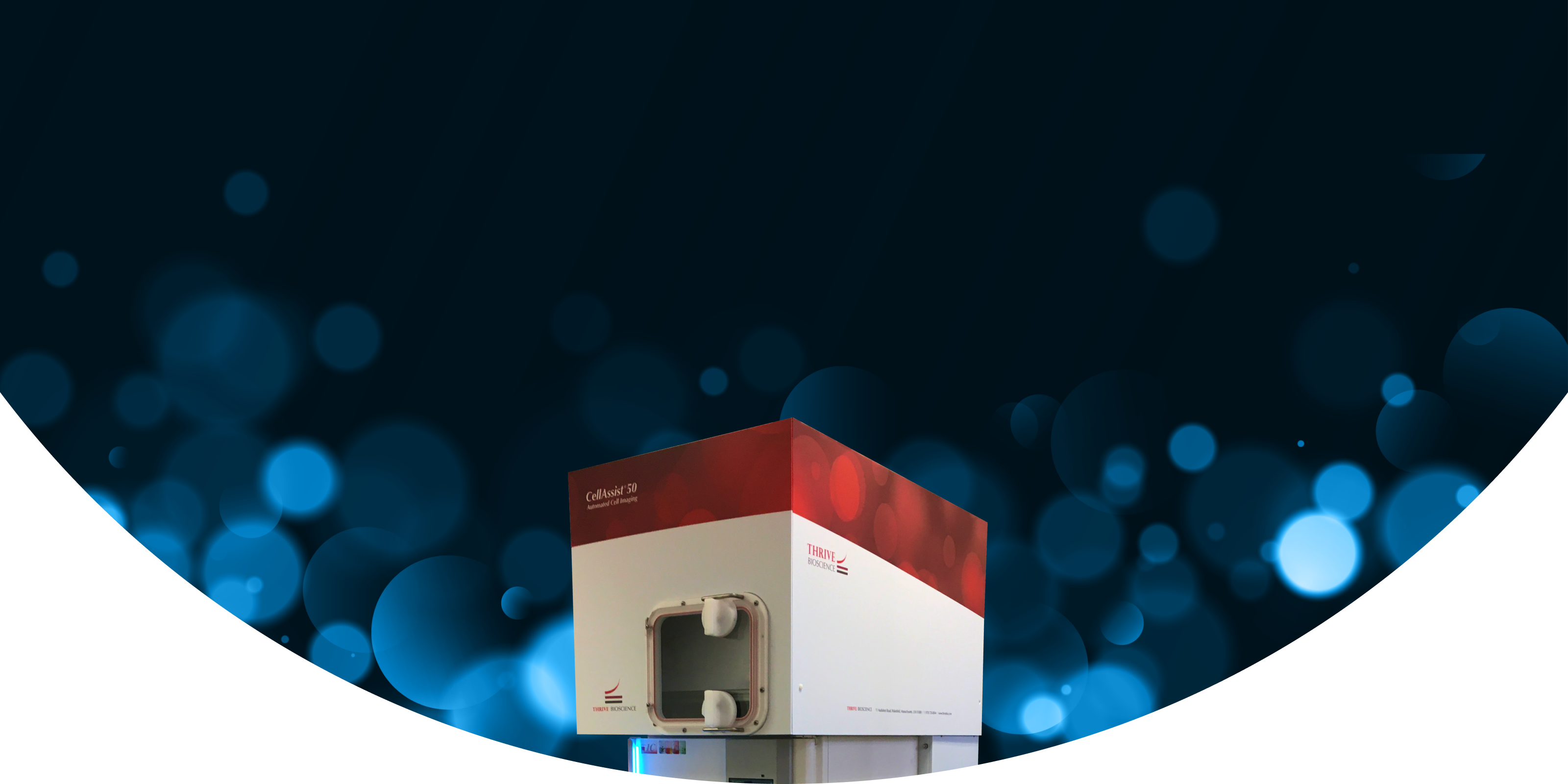
An automated time series cell imaging system with the same imaging and software that drives the CellAssist Benchtop Imager, paired with an integrated 50-plate CO2 incubator. The CellAssist 50 System is ideal for large-scale experiments, or managing multiple projects and users in parallel. With dynamic scheduling, each of the plates can have separate imaging schedules and imaging modes, allowing for consistently timed around-the-clock experiments. Run multiple long-term experiments at the same time, or one large scale a experiment, all with seamless automation.

High-Throughput Efficiency
Image up to 50 cell culture plates automatically with up to 400 scans per day, significantly increasing your data collection capacity and saving you valuable time that would otherwise be spent on manual tasks. Superior image registration allows for enhanced tracking and analysis of live cells frequently over time.

Comprehensive Data Collection
Utilize advanced Thrive quantitative imaging (enhanced and proprietary quantitative phase image) to capture rich, multi-dimensional data across all plates, providing researchers with a deeper understanding and enhancing the quality of your research insights.

Automated Workflow Management
Dynamic scheduling optimizes imaging sequences for multiple users and projects, decreasing operator error while ensuring workflow progresses smoothly and efficiently.

Complete Environmental Control
Precise control over temperature, CO2 (tri-gas option), and humidity ensures an optimal environment for cell health and viability while achieving consistent, high-quality imaging results.

Scalable and Time-Efficient
The CellAssist 50 System automates imaging and data management, empowering you to scale your research efforts while freeing up valuable time for analysis and discovery.
CellAssist 50 System Features
CellAssist 50 System Benefits
Download Brochure
Please fill out the form below for more information about Thrive Bioscience’s technologies.
"*" indicates required fields
Discover how Thrive Bioscience's advanced live cell imaging solutions are transforming research with deeper insights, greater efficiency, and unmatched accuracy across diverse applications.
Explore Applications
Connect with one of our expert staff to discuss how Thrive Bioscience technologies can enhance your cell biology research.
Contact Us
Learn more about our technologies and their use in advancing cell biology research.
RESOURCES
Poster
Using novel, label free intrinsic biomarkers generated from QPI images to gain earlier and deeper insights into microplate cell cultures
October 27th, 2025
View More
RESOURCES
Poster
Cell Proliferation Poster from Gannon University
October 9th, 2025
View More
RESOURCES
Application Note
The CellAssist 50 in BSL-3 Facilities
May 1st, 2025
View More