Contact Us
100 Cummings Center, Suite 407-P, Beverly, Massachusetts 01915, USA
+1 (978) 720-8044
google-site-verification=_llX3wt-_sILNZ4OKcVhHaUFsbcxl34KDwm75iPzM38
Contact Us
100 Cummings Center, Suite 407-P, Beverly, Massachusetts 01915, USA
+1 (978) 720-8044
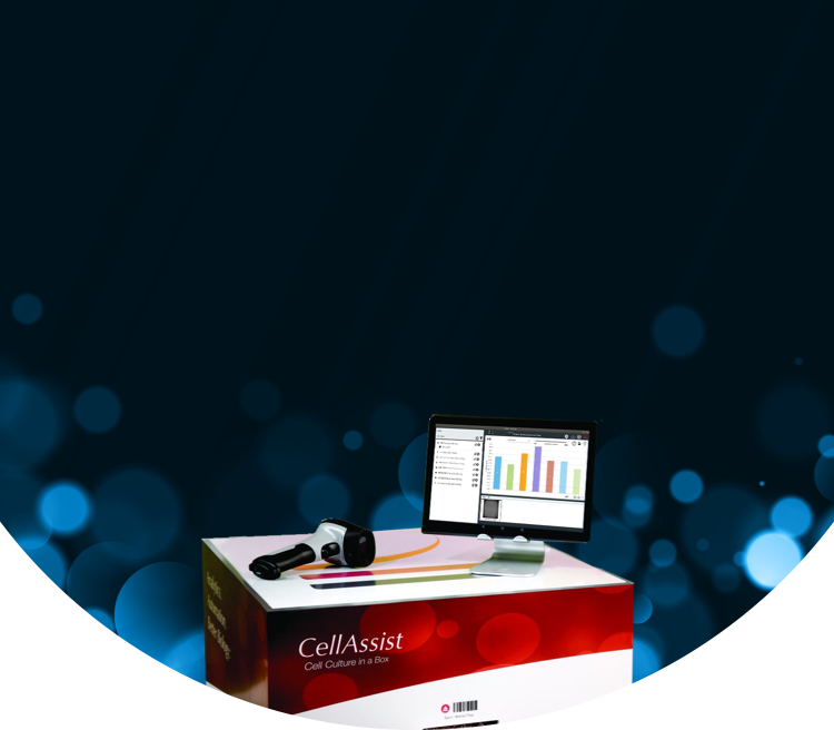
An environmentally-controlled, benchtop system that images single cell culture plates from multiple users. The imaging system provides bright-field, phase contrast, and Thrive Phase Imaging at 4x, 10x, and 20x at 100+ focal planes on 6- well through 384-well flat and round bottom plates. The CellAssist Software Suite automatically builds a centralized database of images, metrics, and methods and materials.
The "CellAssist Experience” video (3:38 minutes) provides an overview of the CellAssist Family of Systems, which combine an advanced imaging system with intuitive software to provide high-resolution images with excellent accuracy. The unique capabilities outlined in the video enable biologists to gain a deeper insight into cell dynamics, and greatly improve the reproducibility of cell, tissue, and suspension culture experiments. Learn more about the CellAssist Family of Systems and their capabilities with this video.

Enhanced Data Quality
Image single plates automatically, significantly increasing your data collection capacity and saving you valuable time that would otherwise be spent on manual tasks. Superior image registration allows for enhanced tracking and analysis of live cells frequently over time.

Remote Data Access
The CellAssist Benchtop Imager provides researchers with real-time access to cell monitoring data, allowing them to work remotely, schedule tasks, and perform analyses from anywhere. The system's 24/7 automation also eliminates the need for overtime, ensuring round-the-clock operation without additional manual effort.

Reproducibility and Traceability
Ensure consistency across experiments with precise data capture and centralized storage, making it easy to reproduce and trace every step of your workflow.

Streamlined Workflows
Minimize hands-on time with fully automated image capture, analysis, and documentation, freeing time to focus on core biological experiments and allowing for 24/7 walkaway time series automation.

User-Friendly Operation
The intuitive interface and easy benchtop integration into existing lab environments make the CellAssist Benchtop Imager a seamless addition to any research setting, ensuring a smooth user experience from setup to data analysis.

Time-Efficient Automation
By automating the entire imaging process, the CellAssist Benchtop Imager significantly reduces manual intervention. This allows researchers to conduct more experiments in less time, enhancing their efficiency and productivity. With a high degree of accuracy and confidence, researchers can focus on their core biological experiments.
CellAssist Benchtop Workflow
Step 1

Capture methods and materials
Step 2

Insert plate into CellAssist Benchtop Imager
Step 3

Capture, view, and transmit to database
Step 4

Remove plate for continued workflow
Step 5

Analyze, compare, and share Images
CellAssist Benchtop Features
CellAssist Benchtop Benefits
Download Brochure
Please fill out the form below for more information about Thrive Bioscience’s technologies.
"*" indicates required fields
Discover how Thrive Bioscience's advanced live cell imaging solutions are transforming research with deeper insights, greater efficiency, and unmatched accuracy across diverse applications.
Explore Applications
Connect with one of our expert staff to discuss how Thrive Bioscience technologies can enhance your cell biology research.
Contact Us
Learn more about our technologies and their use in advancing cell biology research.
RESOURCES
Handbook
The Quantitative Phase Imaging and Morphological Analytics Handbook
April 8th, 2026
View More
RESOURCES
Poster
Using novel, label free intrinsic biomarkers generated from QPI images to gain earlier and deeper insights into microplate cell cultures
October 27th, 2025
View More
RESOURCES
Poster
Cell Proliferation Poster from Gannon University
October 9th, 2025
View More